SML Material
- The SML is made of a copolymer of Hydrophilic and Hydrophobic Acrylic with 25% water content.
- It comes with a UV absorber and is available with an additional blue light filter.
SML Design & Optic
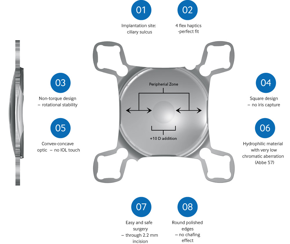
- The SML is an intraocular sulcus lens that has a bifocal design with a central optical zone of 1.5mm in diameter.
- This optical zone provides a +10 diopter addition for patients.
- The peripheral zone is neutral, leaving the patient’s distance vision and visual field unaffected.
- The special convex-concave optic maintains distance between the implants, preventing IOLs from touching eachother.
- Due to its round polished edges the IOL has no chafing effect.
- The IOL’s main square design (patented AddOn technology) prevents iris capture.
- The haptics’ non-torque design ensures rotational stability.
Add-On platform safety
The SML design was based on the Medicontur Add-On Platform design which has gained industry-wide attention for its excellent stability and ergonomic fit within the ciliary sulcus. The Add-On platform has been proven clinically (over 5.000 implantations) and in an in vitro study to be safe.
(Reiter et al. Assessment of a new Hydrophilic Acrylic Supplementary IOL for Sulcus Fixation in Pseudophakic Cadaver Eyes. Eye 2017; 31: 802-809)
MODE OF ACTION
Using the Near Triad Reflex:
miosis – accomodation – convergence
Due to the effect of near vision miosis, the central optical portion that provides the magnified image will dominate when the patient focuses on near objects only, but will not influence far vision significantly when the patient focuses on distant objects through a dilated pupil.
Near vision:
The pupil is constricted (near vision miosis) and light rays pass through the central region of the SML, providing a magnified image on the macula (Figure 1).
Due to the high dioptric power of the central region, sharp vision is achieved at a distance of about 15 cm.

Figure 1:
Patient’s eye with a constricted
pupil (near vision)
Distance vision:
The pupil is dilated when focusing on a distant object and thus light rays passing through the peripheral region of the SML will dominate over those passing through the central region (dashed lines)
(Figure 2).

Figure 2:
Patient’s eye with a dilated pupil
(distance vision)
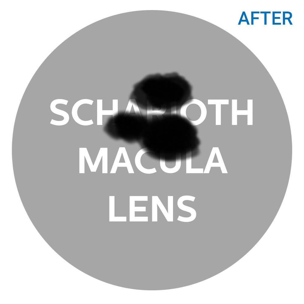
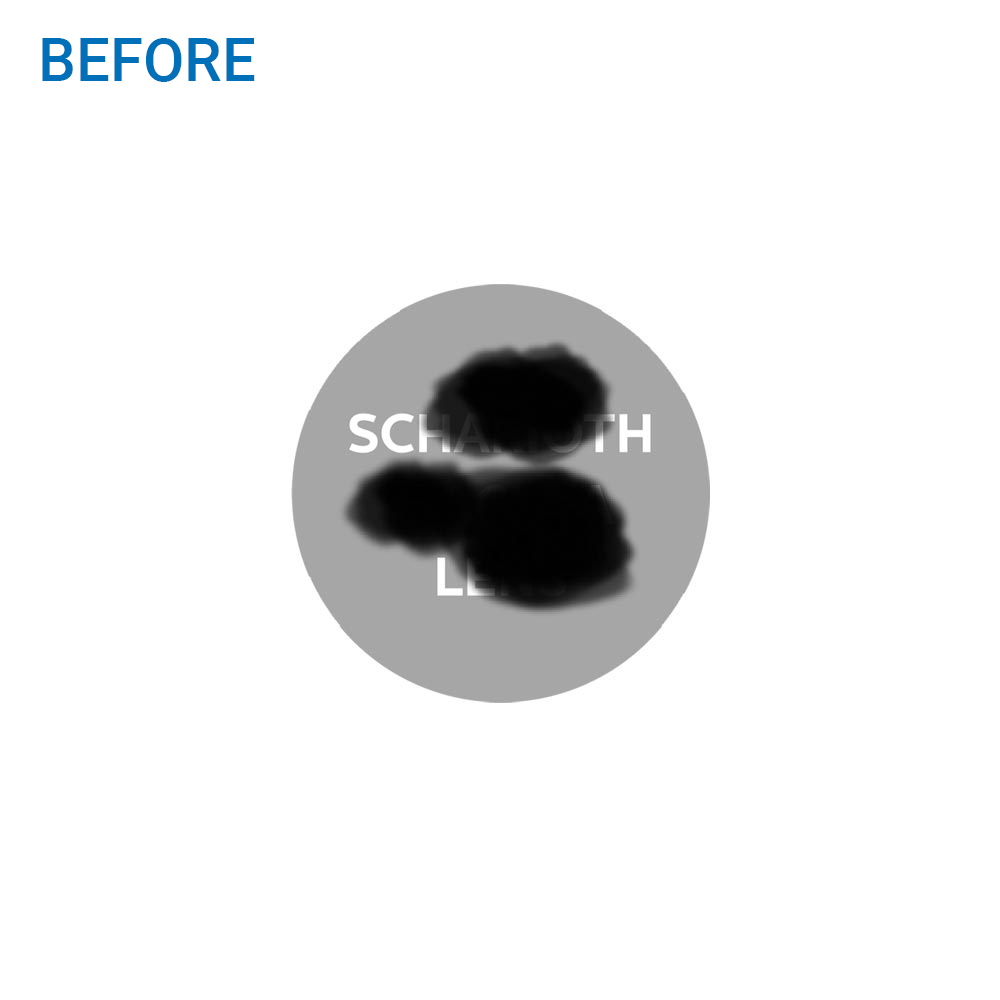
The SML works through magnification (approx. 2.2 times)
The dark spots (scotomas) covering the text represent damaged macular areas.
The SML magnifies the text approximately 2 times but the size of the dark spots remains the same because the SML does not magnify the damaged parts of the macula. Thus, the SML enables patients with dry AMD to read the text.


ADVANTAGES OF THE SML
All examinations needed for a patient’s regular follow-up after implantation with the SML
(retina/macula visualization, OCT etc.) are unaffected.
All treatments, if needed, can be performed without limitations:
- Intravitreal injections in case of exacerbation of maculopathy
- Laser treatment of retina
- YAG capsulotomy
IOL Benefits
- Easy and safe surgery
- Small incision (2.2-2.4mm)
- No visual field reduction
- Unaffected distance vision
- Independent from lens status
- Quick adaptation
- Reversible
- Affordable
PLEASE NOTE:
- SML DOES NOT cure maculopathy.
- SML is a magnifier. It is like a low vision aid inside the eye.
- Exacerbation of maculopathy might occur any time after the implantation of the SML.
- The SML does not affect / limit any diagnostic or treatment procedures for exacerbation

